|
|
 |
|
Image Analysis Systems - Advanced Cytogenetics Imaging - Applied Spectral Imaging - Image Analysis Systems
|
|
|
Model
|
Description
|
|
Products from Applied Spectral Imaging  |
|
HiSKY™
|
High Resolution Spectral Karyotyping Analysis Software
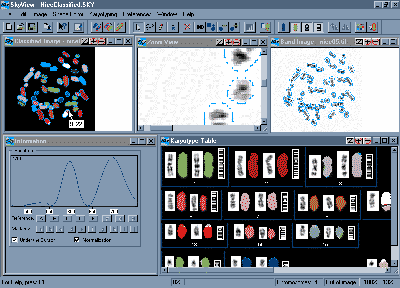
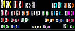
HiSKY was designed to feel similar to SKY, keeping in mind the simplicity and user friendliness that made it the Gold Standard Multicolor FISH application. Existing SKY users will be amazed at how much faster they can obtain more informative HiSKY results. Users new to Multicolor FISH will find HiSKY a must to add to their arsenal of research tools. The HiSKY system offers an integrated solution for your karyotyping needs including BandView and FISHView, which are Applied Spectral Imaging’s band karyotyping and FISH systems.
Quick analysis of the whole genome in one hybridization Accurate detection of chromosomal aberrations Powerful results verification tools Advanced menu organization/ supports customization and simplifies operation View the metaphase and chromosomes in Karyotype table in 8 color options (Enhanced color, band enhanced DAPI, Classified color, and any of the 5 cross-talk-free pure colors used for the staining) Easy switch between the different color image views Enhanced toolbar, improving throughput and simplifying operation Advanced display options providing user-friendly control of all operations Special algorithms for DAPI band-enhancement with customized default Full support for Multi-Species Direct connectivity to Internet sites for immediate reference of relevant aberrations Translocation tool to resolve Even smaller telomeric translocations and insertions Misclassifications in breakpoint regions Mixed classes in overlap or in small chromosomes Advanced Image Enhancement capabilities Ability to perform local DAPI enhancements per selected chromosomes Quantitative Dye intensity information using customized tool tip Full support of DAPI band-based classification (in addition to the spectral classification Advanced Cleaning tool to remove noise while leaving valid translocations Versatile Automatic tools for chromosome definition active in both metaphase and karyotype windows Special Overlap tool for complex overlaps / Allows "painting" each chromosome within the overlap to easily separate the chromosomes Multi-function Cut tool / Separates also overlapping chromosomes Enhanced Join tool / Simply drag and join objects Automatic "Fit to chromosome" tool / Allows boundary expansion or shrinkage for better chromosome contouring Brush Edit tool / Easily erases and adds parts to chromosomes
Multipurpose Magic tool – all-in-one tool / Eliminates the need to switch between tools interprets the mouse movements and performs almost any tool function
Better chromosome alignment with Mirror Image flip tool Recursive Undo operation Ability to display Spine and Centromere of all chromosomes
Download the Datasheet HERE
|
Single Dye Concentration
|
|
IHC Scorer
|
Automatic Immunohistochemistry Scoring
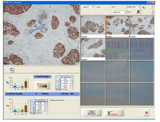
IHC Scorer is Applied Spectral Imaging’s new pathology scoring device.
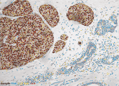
It enables pathologists and researchers to maintain their regular IHC scoring routine andassists them in the determination of the exact final score.
IHC Scorer frees the pathologist from eye assessment of antigen expression
in tissue tumor sections. Due to its unique classification algorithms, it is adaptable to any nuclear or membrane antigen such as ER, PR, Ki-67, p53, Her2Neu and more.
The IHC Scorer brings pathology scoring a big leap forward by allowing
a standard, repeatable, accurate and reliable scoring for better diagnosis,
therapy and laboratory statistics accumulation.
Numerous features allow IHC-Scorer to provide robust answers for expression
levels and cell density.
Download the Datasheet HERE
|
|
|
PathEX
|
Diagnostic tools, Imaging and Information system for the pathology laboratory.
PathEx, the Applied Spectral Imaging suite of diagnostic tools for expert pathologists, brings computer aided technology into a new era of ease and precision.
PathEx tools are designed to assist the pathologists in their everyday tasks
- Laboratory information management
- Determination of diagnostic parameters
- Reports, reviews, confirmation – text and images
- Archiving, statistics
- Transfer to other organization departments
- Connectivity to other organisation departments
The HEART of PathEx – analysis tools - smoothly merge with the pathologist workflow
- Advanced image analysis
- Quantitative diagnostic parameters
PathEx suite applications:
- Immunohistochemistry scoring
- Systematic search for cells or tissue features
- Tissue characterization: staging and grading
- Any other tasks with quantitative measures
Download a Datasheet HERE
|
|
|
AutoMate
|
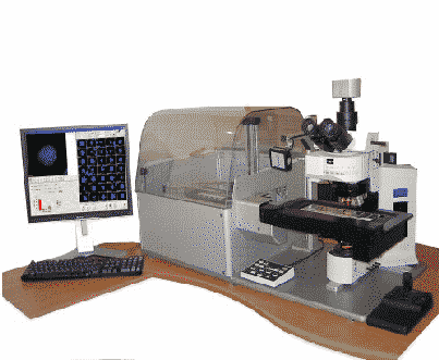
Applied Spectral Imaging's (ASI) has developed the innovative AutoMate Tray Loader to meet the most demanding needs for multi slide scanning,
The AutoMate Tray Loader is available as an add-on to ASI's 9-slide stage ScanView platform.
It allows unattended, continuous scanning of 81 slides
Combination of fluorescent and bright-field scans and tray replacements are fully supported for non-stop operation.
PRODUCT FEATURES
Capacity
- Unattended scan of 81 slides
- Slides are loaded into 9 trays, each containing 9 slides
- Continuous loading/unloading capability to accommodate varying workloads
Functionality
- Barcode reading for automatic case and sample identification
- Optional oil dispenser for unattended object finding followed by high magnification capture using oil objectives
- User definable scan region or multiple regions to match any sample types
- Supports Pre-Scan for definition of regions of interest in tissues
- 9-slide trays to include three cases of triple slide probes
- Non-stop tray replacement designed to go beyond 81 slides
- Desktop design with front loading transparent cover to view trays status
Reliability
- Accurate repeatable tray replacement
- High accuracy in cell-relocation
- Slides are firmly attached in trays
- Manufactured with industry proven and high end components
Flexibility
- Compatible with both Olympus and Zeiss automated microscopes
- Automatic skip on empty slides and trays
- Rapidly review results of any slide in the tray by a simple click of the mouse
- Ability to work with a full set of six objectives
- Ability to combine both fluorescence spot counting slides of various probes with Giemsa stained slides in the same batch
Speed
- Fast loading of trays
- Barcode is read during loading – no extra time needed
- Immediate access for review or relocation to each of the 9-slides in a tray
- Convenient front trays insertion by lab personnel even during scanning
Download a Datasheet HERE
|
|
|
WebView
|
WebView
Web Based Case Sharing
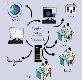
WebView is a web based application that allows cytogeneticists and pathologists to review, respond and remotely analyze cases that are securely shared over the internet.
WebView enables remote review and analysis of cases, captured with all ASI CytoLabView applications; Karyotyping, FISH, CGH, SKY, and Pathex, Pathology applications. It is easy to use, quick to deploy, simple to manage and does not require any special knowledge due to its clear, intuitive interface. It is a secure way to share cases and images across the internet. WebView provides easy and secure remote file access and management 24 hours a day using a simple internet browser.
Download a Datasheet HERE
|
|
|
|
|
|
|
|

