|
|
|
GT Vision News - GT Vision News - GT Vision News - GT Vision News - GT Vision News - GT Vision News
|
|
|
Please select a news topic
|
NEWS TOPIC
|
|
NEW Immunohistochemistry automated scoring from Applied Spectral Imaging
NEW Unique Slide Loader and Scanning System from Applied Spectral Imaging Up to 81 Slides - and its fast!
Filtrex Sales Booming
GT Vision are appointed the exclusive distributors for Applied Spectral Imagings
world-beating Cytogenetics products
New portable imaging System for entomologists
HistoLab histology image analysis over whole tissues
New fast imaging tool for histology and cytology
New filtered particulates analyser for industry
|
NEW, Automatic Immunohistochemistry Scoring

IHC Scorer is Applied Spectral Imagings uniquely powerful and comprehensive new pathology scoring device. It enables pathologists and researchers to maintain their regular IHC scoring routine and assists them in the determination of the exact final score.
|
|
AutoMate Tray Loader
Applied Spectral Imaging's (ASI) has developed the innovative AutoMate Tray Loader to meet the most demanding needs for multi slide scanning,
The AutoMate Tray Loader is available as an add-on to ASI's 9-slide stage ScanView platform.
It allows unattended, continuous scanning of 81 slides. Combination of fluorescent and bright-field scans and tray replacements are fully supported for non-stop operation

|
|
Filtrex Sales Booming
GT Vision USA are reporting record growth in sales of their Filtrex particle analysis systems in the automotive and materials industries.

Filtrex automatically scans, classifies, counts and measures particles trapped in filters. This highly automated system is extremely easy to use and is suitable for all particle sizing applications. With sales running at five times last years record figures GT Vision report that most enquiries arise through existing user referrals giving weight to the great comments from users about the products performance.
|
|
GT Vision are appointed exclusive UK distributors for Applied Spectral imaging
GT Vision today announced that its UK operation have been appointed the exclusive distributor within the UK for all of Applied Spectral Imagings (ASI) automated imaging systems. These products will be supplied through the GT Bio division. ASI is focused on the cytogenetics market with its new EXPO Suite of products. Their products provide high quality imaging solutions for today's clinical and research cytogenetics labs.
ASI offers a complete line of cytogenetics imaging products called CytoLabView , which includes its FDA-approved BandView EXPO™ automated Kariotyping system, its newly-released FISHView EXPO ™ FISH imaging system and SkyView, the gold standard in multicolour Kariotyping. All workstations include a unique manual counting utility and the Case Data Manager (CDM), a powerful and sophisticated patient database. In addition, they have developed software packages such as CGHView , as well as perform a cost-effective multicolour upgrade to SKYView EXPO™ for the most complete clinical cytogenetics imaging package available.
ASI developed and commercialised the gold standard in multicolour Kariotyping (SKYView EXPO™). SKYView EXPO™ simultaneously identifies and displays all chromosomes in unique colours. Its greatest advantage is that it can immediately point out chromosomal aberrations, which are usually an indication of cancer, birth defects or other serious genetic problems, without the need to know the underlying specific disease.
BandView EXPO™, with 510(k) clearance from the FDA, is aimed at the prenatal and oncology clinical markets. FISHView EXPO™ is a general analysis program geared to all fields that use fluorescent imaging, including cytogenetics, pathology, haematology and many others.

ASI also invented SpectraView™, a unique, analytical imaging system that analyses and quantifies information that might otherwise be missed through spectrophotometric analysis of every pixel in an image. This accurate quantification means that the physical and chemical properties of a sample can be quickly and categorically identified, characterised or verified. There are applications for SpectraView™ in drug discovery, genomics, proteomics, cell biology, forensics, entomology, geology, environmental monitoring, histology and pathology.

Ian Baldwin, Sales Director of GT Vision Ltd says, GT Bio is a rapidly expanding division of our company in the fields of scientific imaging, automation and monitoring for life sciences. Applied Spectral Imaging are recognised world leaders in this technology and we expect to significantly expand the number of user in the UK through our expertise in this area and high levels of customer support. The patented, automated multispectral analysis of any object presented to SpectraView™ offers completely unexplored dimensions to the analysis of an enormous range of samples as diverse as paint to insects.
|
|
GT EntoVision - Portable Entomology Imaging System
GT EntoVision launches a whole new way of imaging for the travelling entomologist. Many entomologists frequently travel long distances, either to examine other collections or to collect insects for their own studies. One of the most frustrating difficulties associated with these trips is the often limited facilities available locally for the imaging of specimens. With severe restrictions placed on the movement of specimens by governments and institutions, it is often no longer practical to ship or bring specimens back for examination in their own facilities as may have been done in the past.
As a result of strong demand from the entomological community GT EntoVision have carefully researched and tested a range of components that will now allow entomologists to pack up and take with them their own, high quality, imaging system.
The key aspects of this system are high performance imaging at a reasonable cost, with the additional benefit of being completely portable.
The system includes a fully apochromatically corrected zoom microscope, a scientific grade 3CCD digital camera, a specially selected and configured fibre optic light source, and a powerful, lightweight and portable small form factor computer workstation with GT EntoVisions imaging software all preloaded.
The bulk of the system is housed in a purpose-built shipping case with wheels incorporating cut-outs to cradle all components safely, even in an aircraft hold.
This system represents a significant step forward to entomologists as it includes optics, a digital camera, illumination and software that probably exceed the capabilities of many of the best equipped labs. Now entomologists can be sure of first rate and consistent results no matter where they choose to study their insects.
|
|
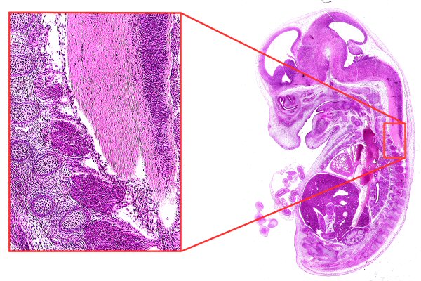
GT HisoVision - Image analysis of large histological features made easy
Trying to extract accurate size and shape information from histological preparations using image analysis can often be difficult, especially when some of the tissues extend beyond the current field of view. Few image analysis products allow features to be joined up across fields. Even fewer manage this using the customers existing microscope stage; nearly always a motorised stage has to be purchased, adding considerably to the cost of the system. This can also significantly deteriorate the users normal interaction with their microscope.
 |
The HistoLab image analysis system from GT HistoVision neatly solves this problem by adding positioning encoders to the existing microscope stage. This tells HistoLab the precise current location on the slide allowing any drawn lines or areas to be continued beyond a single field of view. Additionally, every time a measurement is made a digital picture is taken of the field of view. These pictures and measurements are automatically added to a composite image, giving a superb, high resolution overview of large areas of the slide with all the measured areas included.
HistoLab can be used to compare tissue types, for instance users can count the number of nuclei along a length of intestinal wall or measure the ratio of blood vessel wall thickness to the lumen area. Measurements can be interactive or automatic, including full colour analysis.
|
|
GT Vision Direct - Perfect Color, Cooled Digital Imaging
Scientists engaged in the imaging of histological and cytological specimens use a combination of normal brightfield and fluorescence illumination on their microscope. The difficulty is that very few digital cameras provide live, high resolution colour images as well as enhanced sensitivity cooled chip fluorescence imaging and are therefore optimised for both purposes. The result is the inconvenience and expense of switching between two types of cameras. For instance, it isnt always practical nor possible to take a fluorescence image of a specimen and then combine that image with a colour brightfield picture of exactly the same location in order to obtain some context for the location of the fluorescent markers.
This is just one example of where the new Colour CoolView camera from GT Vision Direct will make a difference. This revolutionary digital firewire camera includes a high resolution 3 CCD sensor, real-time preview at full resolution and peltier cooling. This means that microscope users can have a single camera for both fluorescence and normal brightfield imaging all for the same price as many single chip monochrome cooled cameras.
With its 3 CCDs, Colour CoolView provides perfect colour rendition at the highest possible resolution. The cooling of the 3 CCD head is achieved in a special sealed unit that not only achieves noise reduction due to low temperature but also very quiet electronic noise as the head is isolated from the rest of the cameras built-in image processing electronics.
|
|
GT HistoVision - New tool for Histologists and Cytologists improves specimen imaging quality and speed
Breakthrough technology provided by GT HistoVision called Surveyortm has given routine histologists a new, low cost way to hugely accelerate the process of examining histological sections.
For the first time, Histologists existing microscopes can be upgraded with a motorised imaging system that can scan whole sections at any magnification at the astonishing rate of 25 fields of view per second with no loss of resolution. Simultaneously the images are stitched into one large image of the whole specimen seamlessly. Even more remarkably, simultaneously, the focus is tracked and each field image is stored as well as the ever-growing, large image.
Using the large image as a map, histologists click on any area to immediately obtain, a focused, live image of that field, at any magnification they wish to use. This means that they always have instant access to two magnifications of the sample, and both magnifications can be changed at will.
Users of the system say it reduces the time taken to study sections by up to 95%, virtually eliminates the strain and tedium of microscope work and they automatically have a complete record of every section they wish to keep.
Costing less than many microscopes, the whole system can be installed and working in an hour making it a realistic and practical option for the busy histology lab. If Histologists have an existing camera or motorised stage then GT HistoVision can advise if they can be used or modified to lower the cost of purchase.
GT HistoVision have had a great deal of interest in Surveyortm and are organising several regional seminars throughout the UK where histologists can bring samples and see the system in operation and learn more about the technology deployed.
Surveyortm is developed in the UK and is supported by team of microscope automation and imaging specialists at GT HistoVision.
|
|
GT Vision Direct - Fully Automated Analysis of Filters
It is part of many QC laboratorys daily routine to examine filters under a microscope and to count and characterise the particulates trapped by them. Until now this has been a tedious and very time-consuming task often resulting in only a fraction of the filter being examined and a great deal of operator fatigue and human error. With GT Visions FILTREX system this is a thing of the past. Attaching to existing microscopes or supplied with a new microscope, the FILTREX system allows fully automated, unattended analysis of all or part of every filter using sophisticated image analysis techniques. Analysis times depend on the size of the filter, type of material to be analysed, number and size of particles and proportion of a filter analysed; a typical time is 3 seconds per field. All the operators need to do is load the filter, press start and get on with something else.
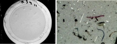 |
For each filter the system generates a report summarising the size distribution and count of particulates per volume of sample with an image of the analysed particles. Perfect for labs needing to validate their test procedures and manufacturing equipment.
Filters are widely used in industries such as pharmaceuticals, cosmetics, foods, beverages, oils, power generation, chemicals, automotive and in environmental air sampling.
Sales Director, Ian Baldwin said ..users of FILTREX have been rather surprised with the improvements in productivity and quality of results generated from day one, with no other sensible alternative, we know that FILTREX will continue to be enthusiastically received by our customers
GT Vision Ltd is the exclusive distributor of FILTREX and a range of other industrial image analysis systems. FILTREX is widely used in Europe especially for component cleaning fluid analysis, hydraulic fluids and pharmaceuticals.
|
|
|