|
|
GT HistoVision |
|
|
GT HistoVision Applications - Histology - Cytology - GT HistoVision Applications
|
|
GT HistoVision Applications |
Application
|
Details
|
|
See how GT HistoVisions products can help you become more productive and produce better results.
rapid slide scanning
ergonomic microscopy
|
Rapid Histology Slide Scanning
|
|
|
This, massive resolution image of a histological section was scanned in less than 1 minute using a x20 objective. The resulting, 1040 fields of view image was stitched into a single image in real-time while the stage was moving using Turboscantm.
After scanning the high resolution map was used side-by-side with a high magnification live image of areas of the section.
In this way two magnifications were made simultaneously available to the operator. By clicking on any region of the map the stage instantly moved to the desired field and automatically refocusing as it moved. The live image of the new field of view at any higher magnification was immediately displayed.
Histologists can make an instant contextural diagnosis without having to switch objectives and move the sample around manually to check locations. Users estimate that this saves several minutes per section.
The images were saved for so that full case notes can be maintained.
|
|
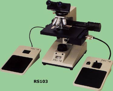
Ergonomic microscopy reduces repetitive strain injury
|
|
|
Users of Ergoscantm significantly reduce the effects of repetitive strains and benefit from improved working conditions. The interface to the operator consists of a sensitive joystick for specimen movement and an ultra-light focus roller.
For routine specimen scanning there is no better, more comfortable way to work. The addition of a high resolution, high speed 3 chip camera and flicker-free display improve operator performance even further and give opportunities to record scenes of interest.
|
|
|